Continuing on previous discussions about complexity in various applications (see previous blog post “About the brain, consciousness and complexity”), here we have applied the concept to a very interesting scenario together with our partners at Helmholtz Zentrum München (HMGU) and at the Institut National de la Santé et de la Recherche Médicale (INSERM) within project Luminous.
The development of the brain… and of consciousness
A question that still puzzles humanity and is still a source of disagreement among experts in various disciplines is whether there is consciousness before birth. When does the magical journey of human consciousness emerge?
In the early stages of development, several cellular processes take place in an effort to form a functional brain and consciousness. During the few months after birth, the brain produces a huge amount of synapses followed by a process of synaptic elimination and stabilization that lasts until adolescence.
Moreover, myelination, a process during which the speed of the electrical pulses’ propagation in the neuronal fibres is increased, starts in the cerebellum and in the brainstem before birth but is completed in the frontal cortex (where executive functions and consciousness reside) in late adolescence. Long-range pyramidal neurons, which are important for conscious processing, are developed around week 26 and indicate that there are already signs of consciousness at this stage.
Measuring foetal brain activity
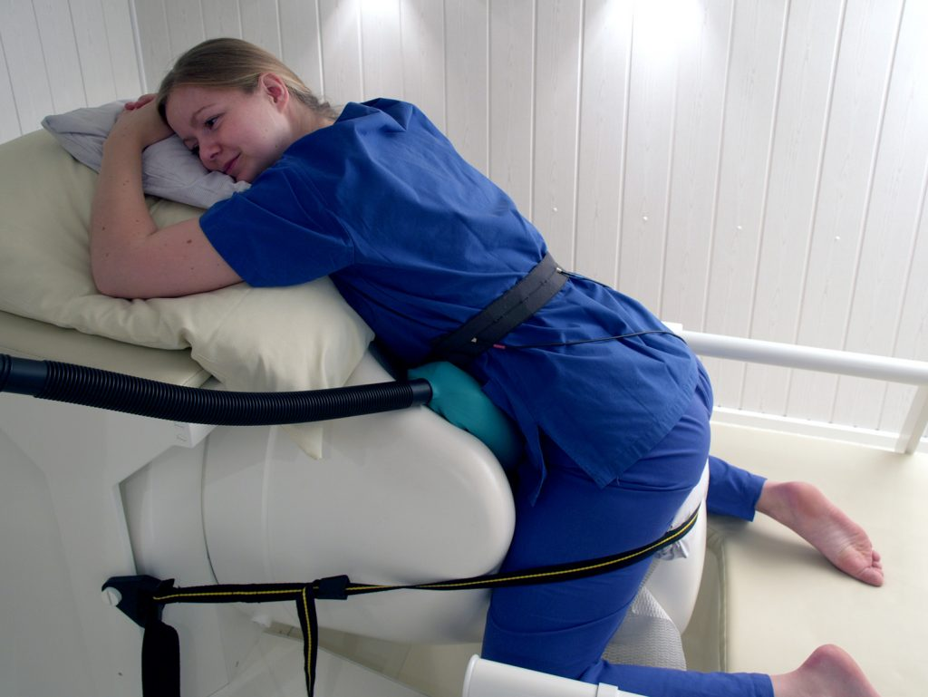
With the current technological breakthroughs, we can now measure the foetal brain activity using foetal magnetoencephalography (fMEG), a tool that in a non-invasive way captures the foetal brain activity in the last trimester of pregnancy and in neonates shortly after birth.
As we already know, brain complexity is related to consciousness, although such a relationship may depend on different measures and theories. Therefore, in order to be able to investigate foetal consciousness, first we need to identify the metrics and theories that best describe our problem.
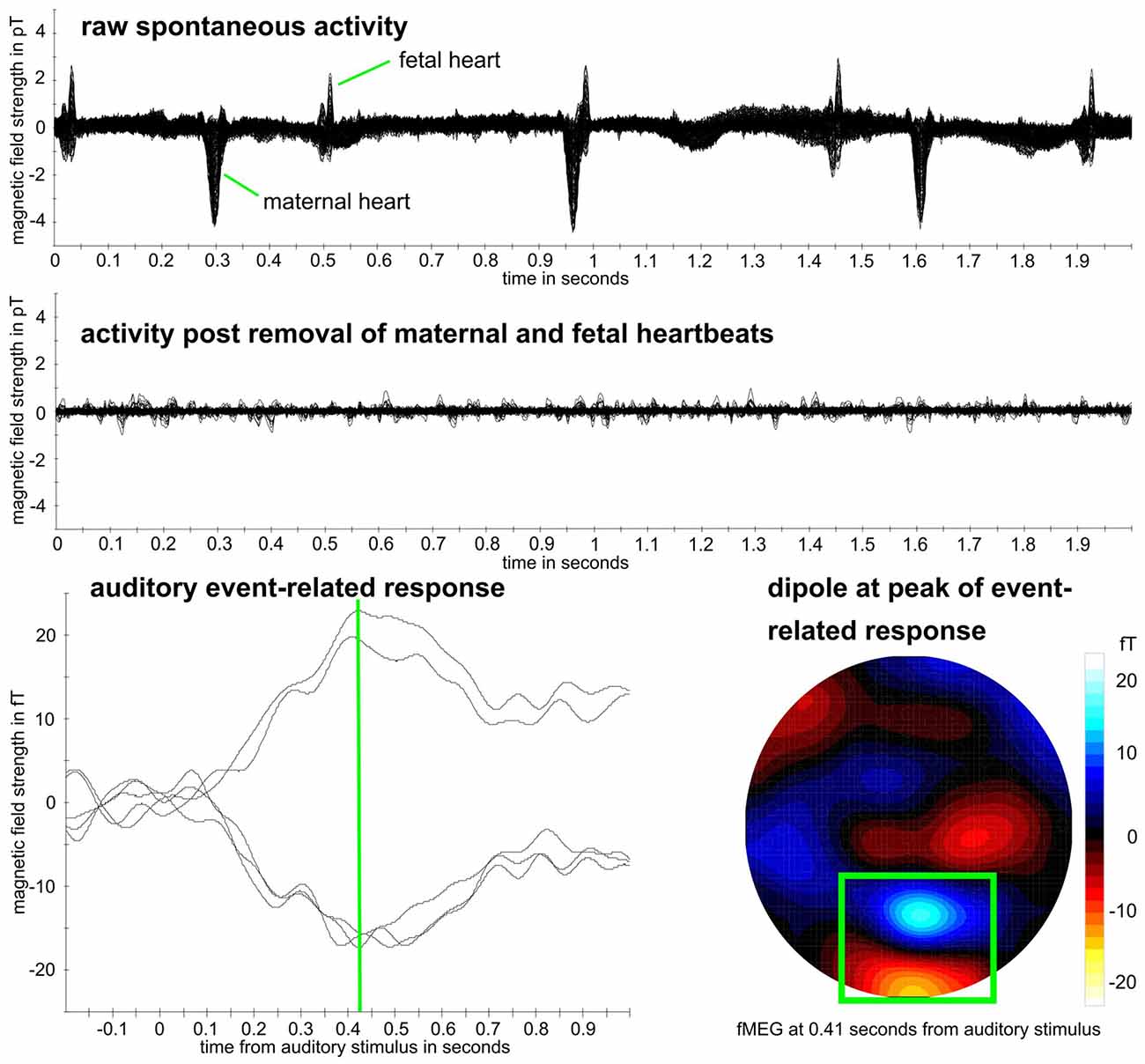
To do this, we acquire and properly clean the recorded fMEG neonatal and foetal data. This involves extracting several elements that describe the complexity of the signals and evaluates them in terms of robustness, efficacy, and speed of computation. This information extraction is done by comparing channels that do not contain any information to channels that contain abundant information.
Eager to go live!
In the Luminous experiment exploring this, the features extracted were derived from two well-established theories on brain complexity: the fractal theory and the compression theory. In the first case, the team extracted measures such as multiscale entropy, multiscale permutation entropy and correlation dimension among others, and in the second case, the Lempel-Ziv complexity measure.
We found that the Lempel-Ziv complexity metric is the most robust one. As well, it is the less computationally complex measure, in contraposition to the rest of the metrics, that require plenty of parameters to be properly defined (making them computationally expensive and robustness-poor).
Having found which metric and theory to use, we are looking forward to seeing how this metric behaves from stage to stage, from foetal to neonatal and to adult brains. What kind of implications for foetal consciousness will it all have? Stay tuned! If you want more details about our study, don’t hesitate to read the whole paper!
This post was originally written by team member Eleni Kroupi, and published at the Neuroelectrics website blog. It has been reproduced here with permission, with some edition for the sake of clarity and adaptation to the format.
Image credits:
fMEG measurement in pregnancy picture, by Luminous team.
Action potential propagation in unmyelinated and myelinated neurons was downloaded from Wikipedia, and licensed via an Attribution 4.0 International (CC BY 4.0) license.
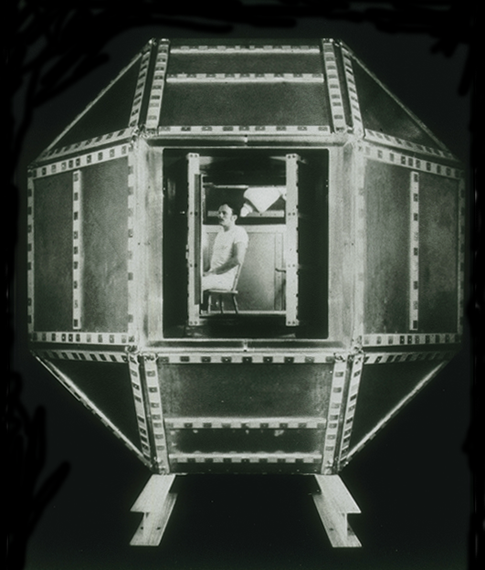
Dr. Cohen’s shielded room at MIT, in which first MEG was measured using SQUID detectors, was downloaded from Wikipedia and licensed via an Attribution-ShareAlike 4.0 International (CC BY-SA 4.0) license.
Example fMEG data was reproduced, via an Attribution 4.0 International (CC BY 4.0) license from the original research article by: Moser J, Bensaid S, Kroupi E, Schleger F, Wendling F, Ruffini G and Preißl H (2019); “Evaluating Complexity of Fetal MEG Signals: A Comparison of Different Metrics and Their Applicability.” Front. Syst. Neurosci. 13:23. doi: 10.3389/fnsys.2019.00023